Common tests and procedures include:

AMSLER GRID
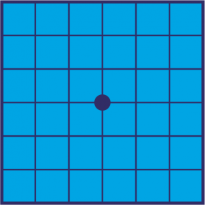
Each eye is checked against an image resembling graph paper with a dot in the center to see if any of the lines appear wavy or missing.

AUTOFLUORESCENCE
Helps determine the area of geographic atrophy in patients with advanced dry AMD.

DILATED EYE EXAM
To view the back of your retina, the doctor dilates the pupils with eye drops.

FUNDOSCOPY OR
OPHTHALMOSCOPY
The pupil is dilated and a bright beam of light is aimed into the eye to view the retina, choroid, blood vessels, and optic disk.

VISUAL ACUITY TEST
Measures ability to see things at different distances.

FUNDUS PHOTOGRAPHY
The doctor uses a customized camera to photograph the back of the eye.

FLUORESCEIN
ANGIOGRAPHY
If the wet form of AMD is suspected, this test may be conducted to detect leaking blood vessels.

OPTICAL COHERENCE
TOMOGRAPHY (OCT)
This technique is used to identify regions of the retina that are thinning, indicating the presence of geographic atrophy.

TONOMETRY
This test measures the pressure inside the eye.